Introduction to Virology
|
Detailed notes for these documents
can be found in Chapter 1 of Principles of Molecular Virology.
|
 Standard
Version: The 4th edition contains new
material on virus structure, virus evolution, zoonoses, bushmeat, SARS
and bioterrorism, CD-ROM with FLASH animations, virtual interactive tutorials
and experiments, self-assessment questions, useful online resources, along
with the glossary, classification of subcellular infectious agents and
history of virology. (Amazon.co.uk) Standard
Version: The 4th edition contains new
material on virus structure, virus evolution, zoonoses, bushmeat, SARS
and bioterrorism, CD-ROM with FLASH animations, virtual interactive tutorials
and experiments, self-assessment questions, useful online resources, along
with the glossary, classification of subcellular infectious agents and
history of virology. (Amazon.co.uk)
|
 Instructors
Version: The 4th edition contains new
material on virus structure, virus evolution, zoonoses, bushmeat, SARS
and bioterrorism, CD-ROM with all the Standard Version content plus all
the figures from the book in electronic form and a PowerPoint slide set
with complete lecture notes to aid in course preparation. (Amazon.co.uk) Instructors
Version: The 4th edition contains new
material on virus structure, virus evolution, zoonoses, bushmeat, SARS
and bioterrorism, CD-ROM with all the Standard Version content plus all
the figures from the book in electronic form and a PowerPoint slide set
with complete lecture notes to aid in course preparation. (Amazon.co.uk)
|
The Origins of Virology
These notes aim to explain:
- The Nature of Viruses - & the problems with studying them
- Discovery of Viruses
- Development of techniques
i.e. how we got here!
Definitions:
Viruses are:
sub-microscopic, obligate intracellular parasites.
- Virus particles are produced from the assembly of pre-formed components, whereas other agents 'grow' from an increase in the integrated sum of their components & reproduce by division.
- Virus particles (virions) themselves do not 'grow' or undergo division.
- Viruses lack the genetic information which encodes apparatus necessary for the generation of metabolic energy or for protein synthesis (ribosomes).
No known virus has the biochemical or genetic potential to generate the energy necessary for driving all biological processes, e.g. macromolecular synthesis. They are therefore absolutely dependent on the host cell for this function.
- Viroids are small (200-400nt), circular RNA molecules with a rod-like secondary structure which possess no capsid or envelope which are associated with certain plant diseases. Their replication strategy like that of viruses - they are obligate intracellular parasites.
- Virusoids are satellite, viroid-like molecules, somewhat larger than viroids (e.g. approximately 1000nt) which are dependent on the presence of virus replication for multiplication (hence 'satellite'), they are packaged into virus capsids as passengers.
- Prions are rather ill-defined infectious agents believed to consist of a single type of protein molecule with no nucleic acid component. Confusion arises from the fact that the prion protein & the gene which encodes it are also found in normal 'uninfected' cells. These agents are associated with diseases such as Creutzfeldt-Jakob disease in humans, scrapie in sheep & bovine spongiform encephalopathy (BSE) in cattle.
For a fun view of the history of virology visit:
|
The Origins of Virology
Ancient peoples were not only aware of the effects of virus
infection, but in some instances also carried out research into the causes &
prevention of virus diseases.
 |
Perhaps the first written record of a virus infection consists of a heiroglyph
from Memphis, the capital of ancient Egypt, drawn in approximately 3700BC,
which depicts a temple priest called Ruma showing typical clinical
signs of paralytic poliomyelitis.
|
|
The Pharaoh Siptah ruled Egypt from 1200-1193 BC when he died
suddenly at the age of about 20. His mummified body laid undisturbed in
his tomb in the Valley of the Kings until 1905 when the tomb was excavated.
The mummy shows that his left leg was withered and his foot was rigidly
extended like a horse's hoof - classic paralytic poliomyelitis.
|
 |
 |
In addition, the Pharoh Ramses V, who died in 1196BC, is believed to have succumbed to smallpox - compare the pustular lesions on the face of the mummy & those of more recent patients. |
 |
 Mummies,
Disease and Ancient Cultures
by Aidan
Cockburn, Eve Cockburn, Theodore A. Reyman (Eds).
Mummies have been found on every continent, some deliberately preserved as with
the ancient Egyptians using a variety of complex techniques, others accidentally
by dry baking heat, intense cold and ice, or by tanning in peat bogs. By examining
these preserved humans, we can get profound insights into the lives, health,
culture and deaths of individuals and populations long gone.
(Amazon.co.UK)
| Smallpox was endemic in China by 1000BC. In response, the practice of variolation was developed. Recognizing that survivors of smallpox outbreaks were protected from subsequent infection, variolation involved inhalation of the dried crusts from smallpox lesions like snuff, or in later modifications, inoculation of the pus from a lesion into a scratch on the forearm of a child. |
 |
 |
On 14th May 1796, Edward Jenner used cowpox-infected material obtained from the hand of Sarah Nemes, a milkmaid from his home village of Berkley in Gloucestershire to successfully vaccinate 8 year old James Phipps.
On 1st July 1796, Jenner challenged the boy by deliberately inoculating him with material from a real case of smallpox !
He did not become infected !!!
To find out more about Edward Jenner & smallpox, click here. |
Although initially controversial, vaccination against smallpox was almost universally
adopted worldwide during the 19th century. Cartoon by James
Gillray, 1802.


Robert Koch (1843-1910) |
However, it was not until Robert Koch & Louis Pasteur jointly proposed the 'germ theory' of disease in the 1880s that the significance of these organisms became apparent. |

Louis Pasteur (1822-1895) |
Koch defined the four famous criteria now known as Koch's postulates which are still generally regarded as the proof that an infectious agent is responsible for a specific disease:
- The agent must be present in every case of the disease.
- The agent must be isolated from the host & grown in vitro.
- The disease must be reproduced when a pure culture of the agent is
inoculated into a healthy susceptible host.
- The same agent must be recovered once again from the experimentally
infected host.
|
Subsequently, Pasteur worked extensively on rabies, which he identified as being caused by a 'virus' (from the Latin for 'poison') but in spite of this, he did not discriminate between bacterial & other agents of disease.

Dmitri Iwanowski (1864-1920) |
On 12th February 1892, Dmitri Iwanowski, a Russian botanist, presented a paper to the St. Petersburg Academy of Science which showed that extracts from diseased tobacco plants could transmit disease to other plants after passage through ceramic filters fine enough to retain the smallest known bacteria. This is generally recognised as the beginning of Virology. Unfortunately, Iwanowski did not fully realize the significance of these results.
|
| A few years later, in 1898, Martinus Beijerinick confirmed
& extended Iwanowski's results on tobacco mosaic virus & was the first
to develop the modern idea of the virus, which he referred to as contagium
vivum fluidum ('soluble living germ'). |

Martinus Beijerinick (1851-1931) |

Freidrich Loeffler (1852-1915) |
Also in 1898, Freidrich Loeffler & Paul Frosch showed that a similar agent was responsible for foot-and-mouth disease in cattle. Thus these new agents caused disease in animals as well as plants. In spite of these findings, there was resistance to the idea that these mysterious agents might have anything to do with human diseases. |
This view was finally dispelled by Landsteiner & Popper (1909), who showed that poliomyelitis was caused by a 'filterable agent' - the first human disease to be recognized as having a viral cause.

Frederick Twort (1877-1950) |
Frederick Twort (in 1915) & Felix d'Herelle (in 1917) were the first to recognize viruses which infect bacteria, which d'Herelle called bacteriophages (eaters of bacteria). In the 1930s & subsequent decades, pioneering virologists such as Luria, Delbruck & many others utilized these viruses as model systems to investigate many aspects of virology, including virus structure, genetics, replication, etc. |

Felix d'Herelle (1873-1949) |
Living Host Systems
In 1881, Louis Pasteur began studies of rabies in animals. Over a number of years, he developed methods of producing attenuated virus preparations by progressively drying the spinal cords of rabbits experimentally infected with the agent, which when inoculated into animals, would protect from challenge with virulent virus. This was the first artificially produced virus vaccine.

Walter Reed (1851-1902) |
During the Spanish-American War of the late 19th century & the subsequent building of the Panama Canal, American deaths due to yellow fever were colossal. The disease also appeared to be spreading slowly northward into the continental United States. Through experimental transmission to mice, in 1900 Walter Reed demonstrated that yellow fever was caused by a virus, spread by mosquitoes. |
| This discovery eventually enabled Max Theiler (1937) to propagate the virus in chick embryos & successfully produced an attenuated vaccine - the 17D strain - which is still in use today.
To find out more about Walter Reed, Max Theiler & Yellow Fever, click here. |

Max Theiler (1899-1972) |
The success of this approach led many other investigators during the 1930s-1950s to develop animal systems to identify & propagate many other pathogenic viruses. Eukaryotic cells can be grown in vitro ('tissue culture') & viruses can be propagated in these cultures, but these techniques are expensive & technically demanding.
| Some viruses will replicate in the living tissues of developing embryonated hens eggs, such as influenza virus. Egg-adapted strains of influenza virus replicate well in eggs & very high virus titres can be obtained. Embryonated eggs were first used to propagate viruses in the early decades of this century. This method has proved to be highly effective for the isolation & culture of many (but not all) viruses. |
 |
 |
In recent years, an entirely new technology has been employed to study the effects on host organisms of viruses: the creation of transgenic animals & plants by means of the insertion into the DNA of the experimental organism of all or part of the virus genome, resulting in expression in the somatic cells (and sometimes in the cells of the germ line) of virus mRNA & proteins. |
Cell Culture Methods:
 |
Cell culture began early this century with whole organ cultures, then progressed to methods involving individual cells, either primary cell cultures (somatic cells from an experimental animal or taken from a human patient which can be maintained for a short period in culture) or immortalized cell lines, which given appropriate conditions, continue to grow in culture indefinitely. |
In 1949, Enders & his colleagues were able to propagate poliovirus in primary human cell cultures.
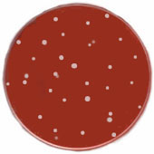
| Renato Dulbecco in 1952 was the first to accurately quantify animal viruses using a plaque assay - dilutions of the virus are used to infect a cultured cell monolayer, which is then covered with soft agar to restrict diffusion of the virus, resulting in localized cell killing & the appearance of plaques after the monolayer is stained. Counting the number of plaques directly determines the number of infectious virus particles applied to the plate. |
 |
Serological/Immunological Methods:
| In 1941 Hirst observed haemagglutination of red blood cells by influenza virus. This proved to be an important tool not only in the study of influenza, but also with several other groups of viruses, e.g. rubella virus.
ONLINE EXPERIMENT: Influenza Haemagglutination |
 |
In the 1960s & subsequent years, many improved detection methods for viruses were developed, for example:
- Complement fixation tests
- Radioimmunoassays
- Immunofluorescence (direct detection of virus antigens in infected cells or tissue)
- Enzyme Linked Immunosorbent Assays (ELISAs)
- Radioimmune precipitation
- Western blot assays
These techniques are sensitive, quick & quantitative.
In 1975, Kohler & Milstein isolated the first monoclonal antibodies from clones of cells selected in vitro to produce an antibody of a single specificity directed against a particular antigenic target. This enabled virologists to look not only at the whole virus, but at specific regions - epitopes - of individual virus antigens. In recent years, this ability has greatly increased our understanding of the function of individual virus proteins. Monoclonal antibodies are also finding increasingly widespread application in other types of serological assay, e.g. ELISAs, to increase their reproducibility, sensitivity & specificity.
Ultrastructural Studies
These can be considered under three areas:
- Physical methods
- Chemical methods
- Electron microscopy
Physical measurements of virus particles began with the earliest determinations of their size by filtration through colloidal membranes with various pore sizes in the 1930s. Experiments of this sort led to the first (rather inaccurate) estimates of the size of virus particles.
The accuracy of these was improved by studies of the sedimentation properties of viruses in ultracentrifuges in the 1960s.
 |
The physical properties of viruses can also be determined by spectroscopy, using both ultra-violet light to examine the nucleic acid content of the particle & visible light to determine its light-scattering properties. |
| Electrophoresis of intact virus particles has yielded some limited information, but electrophoretic analysis of individual virion proteins by gel electrophoresis, & particularly also of nucleic acid genomes, has been far more valuable. |
 |
 |
However, by far the most important method for the elucidation of virus structures has been the study of X-ray diffraction by crystalline forms of purified virus (crystallography). This technique permits determination of the structure of virions at an atomic level. The complete structures of many viruses, representative of many of the major groups, have now been determined at a resolution of a few angstroms. This has advanced our understanding of the functions of the viral particle considerably. |
However, many viruses are resistant to this type of investigation:
- The virus must first be purified without loss of structural integrity. Therefore, adequate quantities of the virus & a method to purify these must be available.
- The purified virus must be able to form crystals big enough to cause significant diffraction of the radiation source.
For some viruses, this is relatively straightforward & crystals big enough to see with the naked eye & which diffract strongly are easily formed. This is particularly true for a number of plant viruses, such as tobacco mosaic virus (TMV) (which was first crystallized by Stanley in the 1935) & turnip yellow mosaic virus (TYMV), the structures of which were among the first to be determined during the 1950s. It is significant that these two viruses represent the two fundamental types of virus particle; helical in the case of TMV & isometric for TYMV.
Nuclear magnetic resonance (NMR) is increasingly being used to determine the atomic structure of all kinds of molecules, including proteins & nucleic acids. The limitation of this method is that only relatively small molecules can be analyzed. At present, the upper size limit for this technique restricts its use to molecules with a molecular weight of less than about 30-40,000 - considerably less that even the smallest virus particles. Nevertheless, this method may well prove to be of great value in the future, certainly for examining isolated viral proteins, if not for intact virions.
Chemical investigation can be used to determine not only the overall composition of viruses & the nature of the nucleic acid which comprises the virus genome, but also the construction of the particle & the way in which individual components relate to each other in the capsid. The reagents used to denature virus capsids can indicate the basis of the stable interactions between its components:
- Proteins bound together by electrostatic interactions can be eluted by addition of ionic salts or alteration of pH.
- Proteins bound by non-ionic, hydrophobic interactions can be eluted by reagents such as urea.
- Proteins which interact with lipid components can be eluted by non-ionic detergents or organic solvents.
In addition to its fundamental structure, progressive denaturation can be used to observe alteration or loss of antigenic sites on the surface of particles & in this way, a picture of the physical state of the particle can be built up.
Proteins exposed on the surface of viruses can be labelled with various compounds (e.g. iodination) to indicate which parts of the protein are exposed & which are protected inside the particle or by lipid membranes.
Cross-linking reagents such as psoralens, or newer synthetic reagents with side arms of specific lengths are used to determine the spatial relationship of proteins & nucleic acids in intact viruses.
1931: Ernst Ruska constructed the first electron microscope. Electron microscopes overcome the fundamental limitation of light microscopes from the virologist's point of view, i.e. the inability to resolve individual virus particles due to physical constraints caused by the wavelength of visible light illumination. During subsequent years, techniques were developed which allowed the direct examination of viruses at magnifications of over 100,000X.
 |
There are two fundamental types of electron microscope, the transmission type (TEM) & scanning electron microscopes (SEM).
Although beautiful images with the appearance of three dimensions are produced by the SEM, for practical investigations of virus structure, the higher magnifications achievable with the TEM have proved to be of most value. |
 |
Two fundamental types of information can be obtained by electron microscopy of viruses:
- the absolute number of virus particles present in any preparation (total count)
- the appearance / structure of the particles
Molecular Biology
Virus infection has often been used to probe the working of 'normal' (i.e. uninfected) cells, e.g. macromolecular synthesis. This is true, for example, of the applications of bacteriophages in bacterial genetics, & also in many instances where eukaryotic viruses have revealed fundamental information about the cell biology & genomic organization of higher organisms.
It is now possible to determine the nucleotide sequences of entire virus genomes,
over a thousand of which have now been completed.
Hybridization:
 |
This nucleic acid-centered technology also allowed advances in detection of viruses & virus infections involving nucleic acid hybridization techniques.
A hybridization probe, labelled to facilitate detection, is allowed to react with a mixture of nucleic acids. The specific interaction of the probe sequence with complementary virus-encoded sequences, to which it binds by hydrogen-bond formation between the complementary base-pairs, reveals the presence of the viral genetic material. |
Because of the essentially digital nature of nucleotide sequences, computers have been found to be the ideal means of storing & processing this information. Moreover, they are capable of predicting, with reasonable accuracy, protein sequences (and detecting other functional regions) from nucleotide sequences.
Vast international databases of nucleotide & protein sequence information
have now been compiled, & these can rapidly be consulted by computers to
compare newly determined sequences with those whose function may have been
studied in great detail. |
 |
Virus Diversity & Classification
Sub-cellular agents present a particular problem for taxonomists. They are too
small to be seen without electron microscopes but very small changes in molecular
structure may give rise to agents with radically different properties.
The International
Committee on Taxonomy of Viruses (ICTV) meets every four years.
"Virus Taxonomy", the 7th report of the ICTV was published in 2000. More
than 1,550 virus species belonging to 3 orders, 56 families, 9 subfamilies and
233 genera are recognized in this report. These well characterized viruses
are probably a small proportion of the total number of viruses which exist. By
the time of the next ICTV meeting, undoubtedly many more viruses will have been
added to this list.
The Origin of Viruses
There are (at least!) three theories which seek to explain the origin of viruses:
- Regressive evolution - viruses are degenerate life-forms which have lost many functions that other organisms possess & have only retained the genetic information essential to their parasitic way of life.
- Cellular origins - viruses are sub-cellular, functional assemblies of macromolecules which have escaped their origins inside cells.
- Independent entities - viruses evolved on a parallel course to cellular organisms from the self-replicating molecules believed to have existed in the primitive prebiotic 'RNA world'.
While each of these theories has its devotees & this subject provokes fierce disagreements, the fact of the matter is that viruses exist, & we are all infected with them. The importance of the origin of viruses is that this issue may have practical implications for virology here & now. Such ideas may allow us to predict the properties & behaviour of new viruses, or to develop new drugs based on what is already known about existing viruses.
SUMMARY:
- Viruses are sub-microscopic, obligate intracellular parasites.
- No known virus has the biochemical or genetic potential to generate the energy necessary for driving all biological processes, e.g. macromolecular synthesis. They are therefore absolutely dependent on the host cell for this function.
- Because of their unique properties, specialized techniques are necessary to grow & study viruses.
|
Detailed notes for these documents
can be found in Chapter 1 of Principles of Molecular Virology.
|
 Standard
Version: The 4th edition contains new
material on virus structure, virus evolution, zoonoses, bushmeat, SARS
and bioterrorism, CD-ROM with FLASH animations, virtual interactive tutorials
and experiments, self-assessment questions, useful online resources, along
with the glossary, classification of subcellular infectious agents and
history of virology. (Amazon.co.uk) Standard
Version: The 4th edition contains new
material on virus structure, virus evolution, zoonoses, bushmeat, SARS
and bioterrorism, CD-ROM with FLASH animations, virtual interactive tutorials
and experiments, self-assessment questions, useful online resources, along
with the glossary, classification of subcellular infectious agents and
history of virology. (Amazon.co.uk)
|
 Instructors
Version: The 4th edition contains new
material on virus structure, virus evolution, zoonoses, bushmeat, SARS
and bioterrorism, CD-ROM with all the Standard Version content plus all
the figures from the book in electronic form and a PowerPoint slide set
with complete lecture notes to aid in course preparation. (Amazon.co.uk) Instructors
Version: The 4th edition contains new
material on virus structure, virus evolution, zoonoses, bushmeat, SARS
and bioterrorism, CD-ROM with all the Standard Version content plus all
the figures from the book in electronic form and a PowerPoint slide set
with complete lecture notes to aid in course preparation. (Amazon.co.uk)
|
© MicrobiologyBytes 2007.
 Standard
Version: The 4th edition contains new
material on virus structure, virus evolution, zoonoses, bushmeat, SARS
and bioterrorism, CD-ROM with FLASH animations, virtual interactive tutorials
and experiments, self-assessment questions, useful online resources, along
with the glossary, classification of subcellular infectious agents and
history of virology. (Amazon.co.uk)
  Instructors
Version: The 4th edition contains new
material on virus structure, virus evolution, zoonoses, bushmeat, SARS
and bioterrorism, CD-ROM with all the Standard Version content plus all
the figures from the book in electronic form and a PowerPoint slide set
with complete lecture notes to aid in course preparation. (Amazon.co.uk)


 Mummies,
Disease and Ancient Cultures


